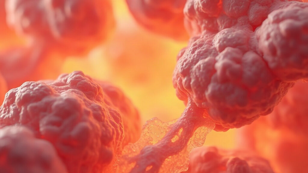
Organ fibrosis is the gradual buildup of collagen-rich scar tissue inside an organ, at the expense of the working cells that keep it functioning. This mechanism affects the liver, lungs, heart and kidneys, and it becomes more common with age. Researchers have established a direct link between the buildup of senescent cells and the progression of fibrosis across several organs. Understanding this process helps people spot early warning signs and act on the levers known to slow it down.
In brief – Organ fibrosis is a biological process that replaces an organ’s functional cells with collagen-rich scar tissue, which stiffens the organ and reduces its capacity to work. It results from a chronic injury (virus, alcohol, smoking, inhaled particles, excess weight) that the body keeps trying to repair without ever completing the healing. With age, senescent cells that accumulate in tissues secrete inflammatory signals that keep this process going, according to work published in Cell and in Nature Communications. Nearly 45% of deaths in developed countries are attributed to a chronic fibrosing disease of the liver, lungs, heart or kidneys, according to a landmark review in the Journal of Pathology. Controlling weight, alcohol, smoking and blood pressure remains the best-documented lever for slowing its progression.
Definition: what is organ fibrosis?
Fibrosis corresponds to an excess deposit of extracellular matrix, mainly collagen, in a tissue that has been repeatedly damaged. According to the landmark review by Thomas Wynn, it results from chronic wound healing that never stops, unlike a normal wound that heals within a few weeks.
The central mechanism runs through the myofibroblast, an activated cell that produces collagen in excess. It comes from several sources:
- Resident mesenchymal cells activated by local inflammation.
- Epithelial or endothelial cells transformed through epithelial-mesenchymal transition.
- Circulating fibrocytes originating from bone marrow.
Once established, this buildup stiffens the organ, hinders cellular exchange and reduces its function, whether that means kidney filtration, gas exchange in the lungs or the heart’s contraction.
The mechanism linking cellular senescence and organ aging
Cellular senescence plays a double role in this process, and that ambivalence explains why it worsens with age. At first, it acts as a brake: activated hepatic stellate cells, the source of scar-forming collagen in the liver, become senescent and are then cleared by NK cells, which limits liver fibrosis (Krizhanovsky et al., 2008, Cell).
With age, this cleanup system runs out of steam. Senescent cells accumulate instead of being cleared and continuously secrete inflammatory molecules, known as the senescence-associated secretory phenotype. A study on idiopathic pulmonary fibrosis showed that the senescent cell burden increases with disease severity, and that their secretome directly promotes collagen production by neighboring fibroblasts (Schafer et al., 2017, Nature Communications).
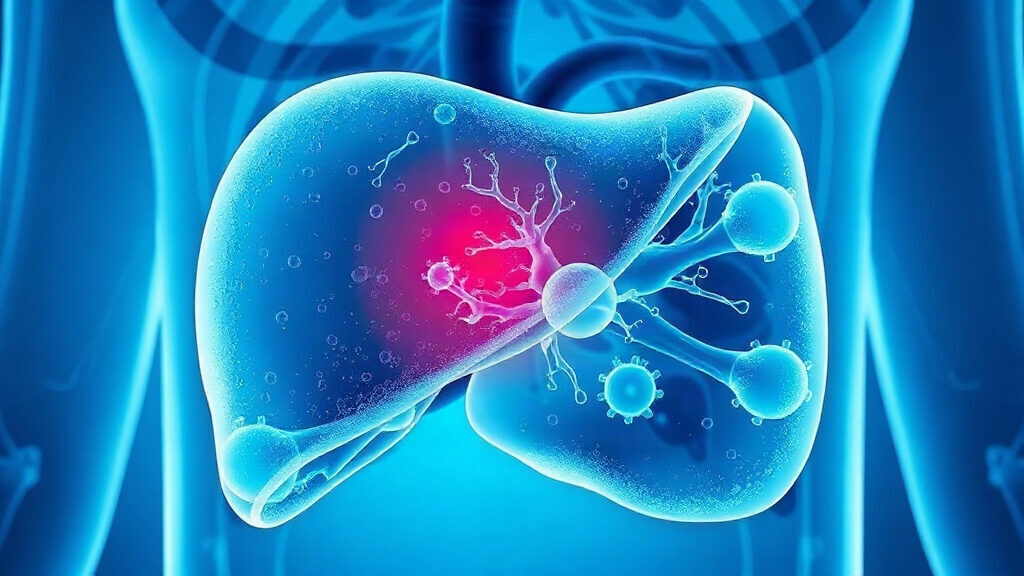
This shift, protective in the short term but harmful when it persists, illustrates why this mechanism is now classified among the processes that speed up organ aging rather than treated as a simple consequence of disease.
What the science says about this progression
A fibrosing disease can affect nearly every organ. According to the landmark 2008 review by Thomas Wynn (Journal of Pathology), nearly 45% of deaths in developed countries are attributed to a chronic fibroproliferative disease of the liver, lungs, heart, kidneys or skin.
In the liver, the National Institute of Diabetes and Digestive and Kidney Diseases notes that fibrosis precedes cirrhosis by several decades of chronic inflammation of the organ. In France, it affects roughly 400,000 people, with 10,000 to 15,000 deaths a year linked to cirrhosis. A reassuring point: if the triggering cause disappears, as when a viral hepatitis is cured, cirrhosis itself can partially regress.
This partial reversibility confirms what cellular senescence researchers observe: the process is not a fixed fate. It depends on the balance between collagen production and the body’s capacity to clear it, a balance that worsens with age but remains modifiable.
In practice: spotting fibrosing damage by organ
Each organ produces different signs, often subtle at first. The table below summarizes the main organs affected, an example of an associated disease and the sign that should prompt a medical visit.
| Organ | Associated disease | Early suggestive sign | Screening test |
|---|---|---|---|
| Liver | Cirrhosis (hepatitis, alcohol, fatty liver) | Persistent fatigue, discomfort under the right ribs | Liver panel, FibroScan |
| Lungs | Idiopathic pulmonary fibrosis | Breathlessness on exertion, dry cough | Chest CT scan, spirometry |
| Heart | Myocardial fibrosis (heart failure) | Breathlessness, swollen ankles | Cardiac MRI, echocardiogram |
| Kidneys | Chronic fibrosing nephropathy | Foamy urine, high blood pressure | Creatinine test, protein in urine screening |
These signs remain nonspecific and shared with other diseases. The example of asbestos and the pulmonary fibrosis it triggers shows how a respiratory injury from decades ago keeps acting long afterward, while alcohol remains one of the main drivers of liver damage in France.
At the cellular level, this progression fits into the broader framework of cellular senescence, one of the recognized mechanisms of biological aging.
Protocol: documented levers for slowing its progression
No treatment currently reliably reverses advanced organ damage in humans, but several levers slow its progression or support partial improvement at early stages.
- Limit alcohol, a direct driver of liver damage and its inflammation.
- Avoid smoking and inhaled particles, which sustain lung damage.
- Treat the underlying cause early (viral hepatitis, diabetes, hypertension) when it is identifiable.
- Monitor weight and insulin resistance, two factors that worsen liver damage.
On this last point, a prospective study of 261 patients with steatohepatitis measured the direct effect of weight loss on repeated liver biopsies: losing at least 10% of body weight was associated with fibrosis regression in 45% of patients (Vilar-Gomez et al., 2015, Gastroenterology). This effect illustrates why weight management remains one of the best-documented levers, without replacing medical follow-up for organs already affected.
Flavonoids such as apigenin, studied for its anti-senescence effects, are the subject of preclinical research on senescent cell burden, though no clinical effect on human disease has been established to date.
Frequently asked questions about organ fibrosis
What exactly is organ fibrosis?
This phenomenon corresponds to the excessive buildup of collagen and other extracellular matrix proteins in a tissue, produced by cells called myofibroblasts. It occurs when an injury (inflammation, virus, alcohol, inhaled particles) repeats itself without giving the organ time to normally repair the damage, which progressively stiffens the affected tissue.
Is organ fibrosis always reversible?
No, reversibility mainly depends on the stage reached. At early stages, removing the triggering cause, such as stopping alcohol or curing hepatitis, can allow partial improvement, as shown by data on cirrhosis. Advanced damage, with significant destruction of the organ’s architecture, rarely regresses completely.
What is the difference between fibrosis and normal wound healing?
Normal wound healing stops once the injury is repaired: the collagen-producing cells disappear through apoptosis or controlled senescence. Fibrosis corresponds to a healing process that never stops, because the injury persists or because the collagen-producing cells escape their normal elimination.
Why does organ fibrosis progress more with age?
The immune system clears senescent cells less efficiently with age, while those cells secrete inflammatory signals that stimulate collagen production in neighboring cells. This buildup, combined with decades of cumulative exposure to alcohol, smoking or excess weight, explains why the process more often takes hold after age 50 or 60.
How can you tell if an organ is affected by fibrosis?
The first signs are often subtle: fatigue, breathlessness on exertion, or localized discomfort depending on the organ involved. Only targeted tests (blood panel, imaging, FibroScan, spirometry depending on the organ) can confirm the diagnosis. Medical advice remains necessary as soon as persistent symptoms appear.
Medical disclaimer. The information provided here is for informational purposes only and does not constitute medical advice. It does not replace a consultation. Ask a healthcare professional before changing your diet, taking dietary supplements or starting a new practice, especially if you have a medical condition, are pregnant or are under treatment. Dietary supplements do not replace a balanced diet or medical follow-up.