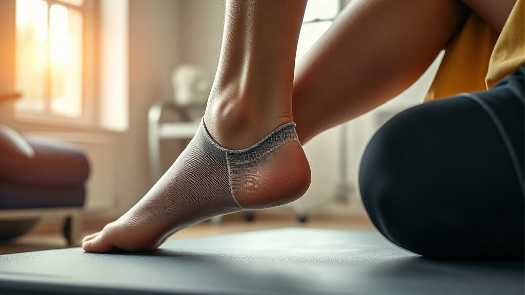
Tendinitis affects the tendon after repeated mechanical overload: a tennis player’s elbow, a runner’s heel, a house painter’s shoulder. Since the 1970s the term has described inflammation of the tendon, but modern imaging and biology tell a more nuanced story, one of successive phases in which inflammation is neither absent nor ever present.
In brief – Tendinitis is a painful tendon condition triggered by repeated mechanical overload, long treated as simple inflammation but now better understood as a continuum of three states: a reactive phase with the release of inflammatory mediators (IL-6, PGE2, substance P), a disrepair phase in which collagen reorganizes poorly, and a degenerative phase in which classic inflammation fades in favor of disorganized tissue. The Achilles, the lateral epicondyle (tennis elbow), the rotator cuff and the patellar tendon are the most affected sites. Prolonged strict rest delays healing: progressive loading, especially eccentric loading, remains the treatment best supported by clinical trials, far ahead of corticosteroid injections, whose benefit reverses after a few weeks.
Definition: tendinitis, tendinopathy or tendinosis?
Medical vocabulary has shifted faster than everyday usage. Tendinitis implies active inflammation of the tendon, with an influx of classic immune cells (neutrophils, then macrophages) as in a fresh sprain. Yet biopsies of chronically painful tendons rarely show this picture: few inflammatory cells, but disorganized collagen, abnormal blood vessels (neovascularization) and nerve endings regrowing into the damaged tissue. This finding has pushed the vocabulary from tendinitis toward tendinopathy, a generic term covering any tendon pain and dysfunction, regardless of how much inflammation is actually present.
Tendinitis nonetheless keeps a precise meaning in one case: the initial, so-called reactive phase that follows an unusual spike in load (returning to sport, moving house, a new racket). At this stage, inflammatory markers are measurable in the tissue, as confirmed by human biopsies published in the American Journal of Sports Medicine. Past this stage, the picture becomes more degenerative than truly inflammatory.
The mechanism: how inflammation takes hold in the tendon
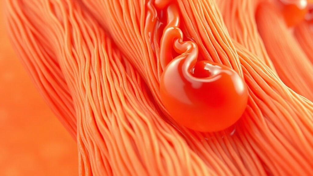
A tendon transmits force from muscle to bone through type I collagen fibers aligned like cables. A sudden or repeated mechanical load beyond the tissue’s capacity to adapt triggers a multi-step biological cascade rather than a single reaction.
- Reactive phase: tenocytes (tendon cells) sense the mechanical stress and release pro-inflammatory cytokines (IL-6, IL-1 beta), prostaglandins (PGE2) and neuropeptides such as substance P, which sensitize nerve endings and account for the early pain.
- Disrepair phase: the tendon attempts to repair its matrix but produces type III collagen, more supple and less resistant, in place of the original type I collagen. Immature blood vessels colonize the area.
- Degenerative phase: after months of unresolved overload, the matrix stays durably disorganized. Classic inflammatory cells become scarce, but macrophages and mast cells persist in small numbers, sustaining low-grade inflammatory signaling without the loud picture of acute inflammation.
This continuum model, proposed by Australian researchers Cook and Purdam, explains why tendinitis can remain painful months after any visible inflammation has disappeared on ultrasound: the pain then comes more from structural disorganization and nerve sensitization than from an influx of immune cells. As the NHS notes, tendon injuries typically follow sudden, sharp movements or repetitive activity such as running, jumping or throwing, which confirms the transient role of inflammation, useful for healing but harmful if it persists (NHS on tendonitis).
What the science says about tendinitis
For a long time, the scientific literature described chronic tendinopathy as a purely degenerative process, without inflammation. A review published in the British Journal of Sports Medicine by Rees, Stride and Scott qualified that view: inflammatory mediators and a discreet immune infiltrate are found even in tendons that have been painful for a long time, reopening the question of inflammation’s role at every stage (Rees et al., 2014).
A human biopsy study led by Millar and colleagues confirmed an infiltrate of mast cells and macrophages as early as the initial stages of tendinopathy, a signal in favor of an innate immune component active earlier than previously thought (Millar et al., 2010). The three-phase continuum model, still used as a reference in sports rehabilitation to tailor treatment to each stage, comes from the work of Cook and Purdam (Cook & Purdam, 2009).
On the treatment side, a meta-analysis of randomized trials published in The Lancet compared corticosteroid injections with other approaches for lateral epicondylitis: clear relief at 4 to 6 weeks, but significantly worse results at one year than simple relative rest or physical therapy (Coombes et al., 2010). Conversely, a progressive eccentric calf-loading protocol allowed 15 patients with chronic Achilles tendinopathy to return to full sports activity after 12 weeks, in one of the founding trials of this approach (Alfredson et al., 1998).
In practice: where and how tendinitis shows up
Four locations account for most tendinitis cases diagnosed in sports medicine and occupational medicine. The Achilles tendon, the lateral epicondyle of the elbow, the shoulder’s rotator cuff and the patellar tendon share the same overload mechanism but different triggers.
| Location | Common name | Frequent cause | Distinctive sign |
|---|---|---|---|
| Achilles tendon | Achilles tendinopathy | Running, abrupt increase in training volume | Pain on waking, stiffness during the first steps |
| Lateral epicondyle | Tennis elbow (epicondylitis) | Repetitive wrist movements, manual tool use | Pain on gripping, on handshake |
| Rotator cuff | Shoulder tendinopathy | Overhead arm movements, carrying loads | Night pain, painful arc on raising the arm |
| Patellar tendon | Jumper’s knee | Repeated jumping and explosive sports | Pain localized under the kneecap during effort |
What these forms of tendinitis have in common: a mechanical load that, session after session, exceeds the tendon’s capacity to regenerate between bouts of stress. Collagen synthesis, which partly depends on protein and vitamin C intake, directly shapes this repair capacity. The Altered intercellular communication pillar groups together several similar inflammatory mechanisms, explored further in our article on chronic inflammation (inflammaging).
Protocol and tips for tendinitis
Managing tendinitis is no longer limited to the rest, ice, anti-inflammatories formula inherited from the 1980s. The tendon needs controlled loading to reorganize, not total immobilization.
- Reduce the load without stopping it: lower the volume or intensity of the causal activity for 1 to 2 weeks, without a complete stop, to avoid letting the tendon deteriorate.
- Introduce progressive eccentric work: the protocol popularized by Alfredson (3 sets of 15 repetitions, twice a day, over several weeks) remains the reference for the Achilles tendon and is adapted to other locations under a physical therapist’s supervision.
- Reserve corticosteroid injections for very painful flare-ups, keeping in mind their unfavorable medium-term effect on tendon structure.
- Correct the movement or equipment behind the tendinitis: adjusting a workstation, changing running shoes, tweaking a racket grip depending on the location.
- Count in months, not weeks: collagen reorganization generally takes 3 to 6 months; improvement that is too slow should prompt a consultation rather than abandoning the protocol.
Resistance training after age 50 illustrates this principle well: progressive, regular loading protects tendons better than prolonged rest, provided recovery phases between sessions are respected.
Frequently asked questions about tendinitis
What exactly is tendinitis?
Tendinitis refers to a painful tendon condition triggered by repeated mechanical overload. The term suggests active inflammation, but research shows that only the initial, reactive phase presents a genuinely inflammatory profile. Past this stage, doctors prefer to speak of tendinopathy, a broader term covering chronic and degenerative tendon conditions.
Does complete rest cure tendinitis?
No, prolonged total rest does not speed healing and can even delay it. The tendon needs controlled mechanical loading to reorganize its collagen fibers. Progressive loading protocols, especially eccentric work, show better results than immobilization in clinical trials on Achilles tendinopathy and epicondylitis.
How long does tendinitis last?
The initial reactive phase can ease within a few weeks if the load is adjusted in time. Tendinopathy that has been present for several months generally requires 3 to 6 months of structured rehabilitation for clear improvement, sometimes longer for the Achilles tendon or the rotator cuff. Consistency in the protocol matters more than its intensity.
Do anti-inflammatories treat tendinitis?
Nonsteroidal anti-inflammatory drugs can reduce pain in the short term during the reactive phase, without addressing the mechanical cause or speeding collagen reorganization. Corticosteroid injections relieve pain quickly but show worse results at one year than physical therapy in several randomized trials, notably for epicondylitis.
When should you see a doctor for tendinitis?
A consultation is recommended if tendinitis pain persists beyond two to three weeks despite load adjustment, if it worsens at night, or if it comes with marked swelling or loss of strength. Medical or physical therapy advice helps rule out a partial tendon tear and adapt the rehabilitation protocol.
Medical disclaimer. The information provided here is for informational purposes only and does not constitute medical advice. It does not replace a consultation. Ask a healthcare professional before changing your diet, taking dietary supplements or starting a new practice, especially if you have a medical condition, are pregnant or are under treatment. Dietary supplements do not replace a balanced diet or medical follow-up.