
Knee cartilage does not wear out like the sole of a shoe: in an uninjured adult, moderate exercise feeds it rather than damaging it. The question matters to anyone who walks, runs, cycles or does strength training after 40 and worries about “wearing out” their joints. MRI studies and large cohorts of runners, published between 1992 and 2019, show that the risk comes mainly from inactivity, injuries, excess weight and extreme training volumes. Here is what effort really does to this tissue, with the figures to back it up.
In brief – Knee cartilage is a tissue without blood vessels that absorbs shocks and feeds itself through compression: each step squeezes it by a few percent, then it refills with joint fluid. A 2017 meta-analysis covering 114,829 people found hip or knee osteoarthritis in 3.5% of recreational runners, compared with 10.2% of sedentary people and 13.3% of competitive runners. A Swedish trial from 2005 even measured an improvement in cartilage composition after 4 months of moderate exercise. The real drivers of wear are knee injuries (cruciate ligament, meniscus), excess weight and very high training volumes sustained over many years. The collagen network barely renews itself after growth ends: protecting the knee from trauma therefore matters as much as staying active.
Knee cartilage: definition and role
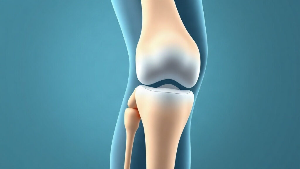
Articular cartilage is a smooth layer a few millimeters thick that covers the end of the femur, the top of the tibia and the back of the kneecap. Its role is twofold: spreading loads over a large surface and letting the bones glide with almost no friction. It contains no blood vessels, no nerves and no lymphatic vessels, which explains both why it does not hurt and why it repairs itself so poorly.
This tissue is built and maintained by a small number of cells, the chondrocytes, scattered through a matrix that they produce themselves. The matrix combines a network of type II collagen fibers, which provides tensile strength, with large molecules called proteoglycans (including aggrecan), which hold water and provide elasticity. Most of the whole structure is water: it is this water, trapped by the proteoglycans, that absorbs the pressure.
One finding changed the way researchers think about wear. In 2016, a Danish team used carbon-14 dating on the cartilage collagen of 23 people born between 1935 and 1997. The result: the collagen network practically stops renewing itself once growth ends, whether the knee is healthy or osteoarthritic (Heinemeier et al., 2016). The framework you have at 20 is, for the most part, the one you will keep. Proteoglycans, on the other hand, are renewed and respond to load.
How exercise acts on knee cartilage: the mechanical pump
Cartilage feeds itself through compression and release, a little like a sponge. With no blood vessels, its oxygen and nutrients come from the synovial fluid, which seeps into the matrix when the load is released. Immobility therefore deprives this tissue of its main source of nourishment.
Felix Eckstein’s team used MRI to measure how kneecap cartilage deformed right after different types of effort in 12 volunteers (Eckstein et al., 2005). Volume fell by 2.8% after walking, 4.5% after cycling, 5.0% after running and 5.9% after knee bends. On the tibial plateau, only landing from jumps (impacts) reached 7%. This compression is temporary: water then flows back into the matrix.
Several mechanisms explain why regular loading keeps the tissue in good shape:
- cyclic compression moves synovial fluid through the matrix and brings glucose and oxygen to the chondrocytes;
- chondrocytes sense mechanical deformation (mechanotransduction) and adjust their production of proteoglycans;
- stronger quadriceps improve shock absorption and reduce pressure peaks on the joint surface;
- movement keeps the joint capsule and the menisci supple, and they spread the load.
Conversely, an excessive or abnormal load (unstable knee, removed meniscus, major excess weight) concentrates pressure on a small area. The chondrocytes then switch to producing enzymes that break down the matrix. With age, some of these cells become senescent: they stop dividing and release inflammatory signals that sustain this breakdown. This link between cellular aging and osteoarthritis is what our pillar page on cellular senescence describes.
What science says about exercise and cartilage wear
The data converge: regular, moderate activity does not damage the cartilage of a healthy knee, and it even seems to maintain it. Risk rises at the two extremes, a sedentary lifestyle and very high training volumes, as well as after an injury.
Cartilage adapts to moderate loading
The first strong signal comes from a Swedish randomized trial. Ewa Roos and Leif Dahlberg followed 45 adults with an average age of 46, who had undergone meniscus surgery 3 to 5 years earlier and were therefore at risk of osteoarthritis (Roos and Dahlberg, 2005). After 4 months of supervised exercise 3 times a week, a specialized MRI technique (dGEMRIC) showed improved glycosaminoglycan content: +15 ms versus -15 ms in the control group. The more participants had increased their activity, the richer this tissue became.
An Australian cohort of 297 adults aged 50 to 79 with no knee injury points in the same direction. Those who did vigorous activity (enough to sweat or get out of breath) had a larger tibial cartilage volume and fewer defects on MRI (Racunica et al., 2007). Regular walking was associated with fewer lesions in the bone beneath the cartilage. This is an association, not proof of cause.
In animals, the relationship seems to follow a U-shaped curve. A review of 29 randomized trials shows that at moderate doses, all 12 comparisons report a neutral or positive effect on the matrix, whereas at high doses, 19 of 27 comparisons report degradation (Bricca et al., 2017). In humans who already have osteoarthritis, a review of 9 trials concludes that weight-bearing exercise does not appear harmful to cartilage, with low-quality evidence (Bricca et al., 2019).
Running: less osteoarthritis in recreational runners
Running is the sport most suspected of wearing out knees, and it is also the best studied. The Alentorn-Geli meta-analysis pools 25 studies and 125,810 people (Alentorn-Geli et al., 2017). The prevalence of hip or knee osteoarthritis was 3.5% in recreational runners, 10.2% in sedentary people and 13.3% in competitive runners. The authors stress that they cannot rule out bias, particularly from previous injuries.
In the American Osteoarthritis Initiative, 2,637 adults with an average age of 64 described their sporting history in detail. Having run did not increase the risk of symptomatic knee osteoarthritis; among current runners, the crude odds ratio was 0.64 (Lo et al., 2017). A second analysis of 1,203 people who already had osteoarthritis found no radiographic worsening linked to running over 4 years (Lo et al., 2018).
The marathon adds a useful nuance. In 82 middle-aged first-time marathoners followed by MRI, bone marrow edema beneath the cartilage of the tibia and femur decreased after the race, but the cartilage on the outer side of the kneecap deteriorated slightly, without symptoms (Horga et al., 2019). The knee therefore responds differently from one zone to another, and a large volume packed into a few months mainly stresses the kneecap joint.
Injuries and excess weight: the real accelerators of knee cartilage wear
An anterior cruciate ligament tear weighs far more heavily than years of jogging. Twelve years after this injury, 51% of 67 Swedish female soccer players had radiographic knee osteoarthritis, at an average age of 31 (Lohmander et al., 2004). Neuromuscular warm-up programs halve the risk of cruciate ligament tears, and cut non-contact tears by two thirds in female athletes (Webster and Hewett, 2018).
Weight acts as a multiplier. In 142 adults with osteoarthritis, each kilogram lost reduced the force transmitted to the knee at every step by about 4 kg, or roughly 4 lb per pound lost (Messier et al., 2005). In the Framingham study, women who had lost about 5.1 kg (11.2 lb) over 10 years saw their risk of symptomatic knee osteoarthritis cut in half (Felson et al., 1992). MedlinePlus, the US National Library of Medicine’s health service, also lists excess weight and joint injuries, including those from sports, among the risk factors for osteoarthritis (MedlinePlus page on osteoarthritis).
| Study | Population | Main finding |
|---|---|---|
| Heinemeier, 2016 | 23 people, carbon-14 dating | Cartilage collagen almost not renewed after growth |
| Eckstein, 2005 | 12 volunteers, MRI before and after exercise | Temporary compression from 2.8% (walking) to 5.9% (knee bends) |
| Roos and Dahlberg, 2005 | 45 adults after meniscus surgery | Better cartilage composition after 4 months of exercise |
| Racunica, 2007 | 297 adults aged 50 to 79 | Vigorous activity associated with greater cartilage volume |
| Alentorn-Geli, 2017 | 125,810 people, 25 studies | Osteoarthritis: 3.5% (recreational), 10.2% (sedentary), 13.3% (competitive) |
| Lohmander, 2004 | 67 female soccer players, 12 years after a cruciate ligament tear | 51% radiographic osteoarthritis at around age 31 |
In practice: which activities keep knee cartilage healthy?
The best activity for knee cartilage is the one you do regularly, at a moderate load, without pain that lingers the next day. The knee tolerates variety very well, and combining endurance exercise with strength training covers most needs. The WHO points out that exercise and keeping a healthy weight are core tools against osteoarthritis, which affected 528 million people worldwide in 2019, including 365 million in the knee (WHO fact sheet on osteoarthritis).
Cycling puts moderate compression on the kneecap and almost no impact on the tibial plateau, which makes it a safe foundation (see our article on the effects of cycling on the heart and knees). Running remains compatible with a healthy knee if volume increases slowly. Strengthening the thighs protects the joint by absorbing part of the impact: our guide to strength training after 50 details suitable exercises.
| Activity | Load on cartilage | Strength | Precaution |
|---|---|---|---|
| Walking | Low (kneecap compression 2.8%) | Accessible at any age, associated with fewer bone lesions | Cushioned shoes if overweight |
| Cycling | Moderate, no impact | Moves the knee without shock, ideal when returning to exercise | Saddle height set to avoid excessive bending |
| Recreational running | Moderate to high, with impacts | Osteoarthritis less common than in sedentary people | Gradual increase in volume, varied surfaces |
| Swimming, water aerobics | Very low | Useful when the knee is already painful | Limit breaststroke if the knee is sensitive |
| Strength training | Moderate, controlled | Stronger quadriceps, better shock absorption | Gradually increase range of motion in squats |
| Pivoting sports (soccer, skiing) | High, twisting | Endurance and coordination | Risk of cruciate ligament and meniscus injury |
When osteoarthritis has already set in, exercise remains fully relevant. The 2015 Cochrane review, which pools 44 trials and 3,537 participants, shows a 12-point reduction in pain on a 100-point scale and a 10-point gain in function, an effect comparable to that of anti-inflammatory drugs (Fransen et al., 2015). Our article on osteoarthritis and longevity explores this link with healthy life expectancy.
Protecting your knee cartilage: practical tips
Protecting knee cartilage comes down to loading it often, avoiding injuries and limiting excess weight. These steps are based on the studies cited above and suit an adult with no known knee condition.
- Move on most days: 150 to 300 minutes of moderate activity per week, following WHO guidelines, split into shorter sessions if needed.
- Increase your running or brisk walking volume gradually, without doubling the distance from one week to the next.
- Do 2 strength sessions for the thighs and hips per week (partial squats, step-ups, glute bridges).
- Before pivoting sports, do a neuromuscular warm-up: jumps with controlled landings, single-leg balance, changes of direction.
- If you are overweight, even a modest loss relieves the knee: about 4 kg less load per kilogram lost (4 lb per pound), at every step.
- After a knee sprain with rapid swelling, have the joint examined before returning to sport.
Pain serves as a guide. Mild discomfort during exercise that disappears within 24 hours is acceptable; pain that gets worse the next day, swelling or a knee that locks means you should reduce the load and see a doctor. To receive our weekly summaries on the science of joint and muscle aging, sign up for our newsletter.
Frequently asked questions about knee cartilage and exercise
Does running wear out knee cartilage?
In an uninjured adult, recreational running does not wear out knee cartilage. A 2017 meta-analysis of 114,829 people found osteoarthritis in 3.5% of recreational runners versus 10.2% of sedentary people. Risk rises mainly with high-level competition, previous injuries and excess weight.
Can knee cartilage regenerate?
The collagen network of cartilage barely renews itself after growth ends, according to carbon-14 dating published in 2016. Proteoglycans, which hold water, can however become more abundant: a Swedish trial from 2005 measured this by MRI after 4 months of moderate exercise. A deep lesion, by contrast, does not fill in on its own.
Which sport should you choose when your knees hurt?
Cycling, swimming, water aerobics and walking are the best tolerated when the knee is painful, because they work the joint without violent impact. Quadriceps strengthening complements these activities. According to the 2015 Cochrane review, exercise reduces pain by 12 points out of 100 in people with knee osteoarthritis.
Why is immobility bad for cartilage?
Cartilage has no blood vessels: it receives oxygen and nutrients from the synovial fluid, which enters its matrix during cycles of compression and release. Without movement, this pump stops and the cartilage cells receive less nourishment. In large cohorts, sedentary people also have more osteoarthritis than recreational runners.
Does losing weight protect knee cartilage?
Yes, weight loss clearly reduces the load on knee cartilage. In 142 adults with osteoarthritis, each kilogram lost reduced the force transmitted to the knee at every step by about 4 kg. In the Framingham study, a loss of about 5 kg (11 lb) over 10 years halved the risk of symptomatic osteoarthritis in women.
Medical disclaimer. The information provided here is for informational purposes only and does not constitute medical advice. It does not replace a consultation. If you have persistent knee pain, swelling, locking or instability, or after an injury, see a doctor or a physical therapist before resuming or stepping up a sports activity. Ask a healthcare professional before starting a new practice, changing your diet or taking dietary supplements, especially if you have known osteoarthritis, a medical condition, are pregnant or are under treatment. Dietary supplements do not replace a balanced diet or medical follow-up.